How We Hear
 The human ear can be divided functionally into 3 sections: the outer, middle and inner ear. All three parts work together to conduct sounds from our listening environment to the brain for processing.
The Outer Ear
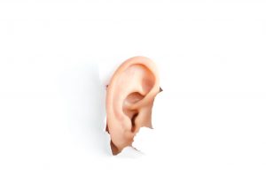
The external ear has two parts: the PINNA which is the outside portion of the ear that is visible on the side of the head, and the EAR CANAL which extends from the pinna to the TYMPANIC MEMBRANE (eardrum). The pinna is mostly skin and cartilage and a few muscular attachments. The pinna collects and directs sounds down the ear canal. The twists and folds of the pinna enhance high pitched sounds and also enable us to determine the location of the sound source, providing better ability to hear from the front and sides compared to sounds behind us. Cupping the hand behind the pinna can provide slight amplification to sounds coming from the front because it effectively enlarges the sound collection surface area. The ear canal is a small, tunnel-like tube that connects the pinna to the eardrum. The outer two thirds of the canal is cartilaginous and contains glands that produce CERUMEN (earwax), while the inner one third is surrounded by bone.
The Middle Ear
The middle ear consists of an air-filled space between the eardrum and inner ear that contains a chain of three OSSICLES (tiny bones) linked together via tiny ligaments and muscles that support and adjust tension as required to conduct sounds. The eardrum is a concave shaped layer of membrane at the end of the ear canal. Sounds travel down the ear canal and strike the eardrum, causing it to vibrate. These vibrations are then transferred through the ossicles to the snailed-shaped organ known as the COCHLEA (inner ear). The bones are known as the MALLEUS, INCUS and STAPES (also known as the hammer, anvil and stirrup). Sound sets this whole structure into vibration and the stapes vibrates into an opening in the cochlea, transferring sound energy into the fluids and tissues of the cochlea.
There is a small tube that connects the middle ear space to the back of the throat known as the EUSTACHIAN TUBE. This tube is normally closed but opens momentarily upon yawning or swallowing. This periodic opening maintains equalization of the air pressure between the middle ear and outside air pressure. This pressure must be equalized for most effective transfer of sounds through the middle ear. If it becomes unequal as with rapid altitude change (i.e. on an airplane) or with a cold, the sudden opening of the Eustachian tube produces a ‘pop’ along with improved hearing because the pressure balance has been restored. Children have smaller, less efficiently positioned Eustachian tubes compared to adults, and this contributes to a higher incidence of blocked ears and subsequent ear infections.
The Inner Ear
The inner ear is comprised of two functionally separate sections: the VESTIBULAR (balance) portion and the COCHLEA (hearing portion). These two are interconnected and each serves it’s own vital function.
The vestibular portion enables us to sense motion and head position in relation to gravity. This area makes it possible to maintain sharp visual focus with the many small and rapid motions of the head that occur as we engage in walking or other activities.
The cochlea is a coiled canal within the dense bone tissue of the skull. Resembling a snail’s shell, it houses three fluid-filled membraneous canals extending it’s full length. The central canal houses the ORGAN OF CORTI, which is comprised of specialized cells and their supporting tissues. Vibratory energy propagated through the fluid produces deformation of the organ of Corti, in turn resulting in shearing forces on the tiny hair-like projections it contains. This shearing action triggers an electro-chemical signal that travels through the auditory nervous pathway which then projects to the brainstem and upward to the auditory processing centres of the brain where we ultimately ‘make sense’ of what we have heard.
This is a simplified description of a complex activity that ultimately results in what we know as ‘hearing’.
Note: the original post can be found here.
About the Author:
 Tina Stafferton is the primary audiologist at Sound Hearing Care. Tina has been providing hearing healthcare to all ages in the Windsor and Essex county area since 1999. Tina has worked extensively in both hospital and private settings. She maintains close relationships with all her patients and prides herself on flexible, individualized care with an emphasis on proven technology.
Tina is a member in good standing with the College of Audiologists and Speech-Language Pathologists of Ontario (CASLPO) and is a registered provider with the Assistive Devices Program (ADP). Tina regularly attends conferences and workshops to keep abreast of the rapidly changing technology and research in the areas of hearing and amplification.
M.S. Audiology, Wayne State University (1999)
Doctor of Audiology (Au.D) Wayne State University (2006)
She owns a clinic in Tecumseh and Belle River. To learn more, please visit her website www.soundhearingcare.ca
